Case Study: Implant replacement of the upper left central incisor utilising MinerOss® and NovoMatrix™

Patient presentation
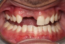
Mrs E.N. was referred for implant replacement of her failed UL1. The tooth was tender to percussion and exhibited grade ii mobility.
Clinical and CT scan assessment confirmed that the UL1 suffered from root resorption and was associated with a large periapical lesion which extended palatally and laterally towards the UL2 root tip. The UL2 tested positive to vitality testing.
Treatment options
It was considered high risk to attempt an immediate implant placement in such a compromised site. The treatment plan was to firstly remove the tooth and the granulation tissue and perform a ridge preservation procedure simultaneously to combat the likelihood of severe shrinkage of the socket during the healing period. The rationale being to fill the bone defect with a bone graft material immediately after tooth removal, to reduce the shrinkage of the socket and maintain most of the socket dimensions during the healing phase.


Treatment: Bone augmentation
The first treatment phase included the elevation of a large full thickness flap to fully expose the area and enable the harvesting of autogenous bone.
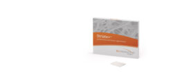
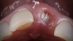
The UL1 was then removed including excavation of all granulation tissue taking care to avoid any direct curettage of the exposed UL2 root tip. The apical defect as well as the UL1 socket was then filled with a 50/50 mix of autogenous bone chips and an allograft (MinerOss® – cortico-cancellous mix) and the apical defect was covered with a native porcine collagen membrane with a high percentage of collagen type iii for a prolonged barrier function of six months (Jason® membrane).
The muco-periosteal flap was sutured back with 6.0 prolene and no attempt was made to coronally advance the flap for complete closure. The rationale was to maintain the open socket wound and thereby preserve the original mucogingival relationship with no alteration. The socket entrance was sealed with a resorbable haemostatic sponge (hemocollagene) and the original tooth was utilised as a temporary pontic and bonded to the adjacent teeth with flowable composite.


Treatment: Implant surgery
Healing was uneventful and following a seven-month healing period, a full thickness flap was elevated revealing good turnover of bone graft into host bone. A bone level implant was placed in a slightly buccal position due to the close proximity of the naso-palatine foramen followed by the placement of a 4mm healing cap. No additional bone grafting was performed but the soft tissue volume was enhanced utilising a strip of acellular porcine dermal matrix (NovoMatrix™) which was sutured to the palatal flap and stabilised buccally with periosteal suture fixation. Full closure was performed with 6.0 prolene.


Three months later a small crestal incision was performed to expose and remove the healing cap and impressions taken to construct an under-contoured screw-retained provisional crown. Over a period of two visits, composite was incrementally added to the subgingival portion of the temporary crown to expand and modify the soft tissue emergence profile until an acceptable shape was achieved. A new impression was taken capturing the transmucosal shape and a customised abutment and zirconia crown was fabricated as a one-piece screw-retained restoration.
Due to multiple connections/disconnections during the restorative stages, epithelial downgrowth had occurred within the transmucosal compartment. Therefore, prior to fitting of the final restoration, the internal aspect of the soft tissues was de-epithelialised (refreshed) with a low grit rugby shaped diamond bur and the final crown fitted immediately afterwards.


Conclusion
A two-month review of the final outcome displayed adequate hard and soft tissue volume at this very early stage of maturation which is likely to improve over the following twelve months. The UL2 maintained vitality throughout the treatment.


Find out more about our biologics and implant solutions here, or contact your BioHorizons Camlog Territory Manager.
Article feedback
Article Author
Rob qualified at King's College, London in 1987. After working in an orthodontic practice in Colchester for four years he moved to Newbury, Berkshire. Rob then spent the next 14 years working in a busy private practice where he built a reputation for treating complex cases involving cosmetic, implant and orthodontic treatments. Rob now spends his time between treating referred patients at Pentangle Dental Transformations and teaching. Rob is a postgraduate tutor for the Thames Valley Deanery, a mentor and faculty educator for the ADI (Association of Dental Implantology), a mentor and international speaker for the ITI (International Team of Implantologists), a Royal College of Surgeons examiner for the implant diploma (RCS Edin) and the Membership in Advanced General Dental Surgery (MAGDS RCS Edin) and was the president of BAAD (British Academy of Aesthetic Dentistry) 2018-2020.