CPD Article – Leukocyte and platelet-rich fibrin, the natural choice
The aim of this article is to present the science behind platelet concentrates with a particular focus on Leukocyte- and Platelet- Rich Fibrin, why it is so important in implant dentistry, and how it works in practice
Aims & Objectives
- Understand the importance of leukocytes and platelet concentrates in implant dentistry
- Understand the difference between Platelet-Rich Plasma, Plasma Rich in Growth Factors, Platelet-Rich Fibrin and Leukocyte- and Platelet-Rich Fibrin
- Understand the evidence base supporting the use and benefits of Leukocyte- and Platelet-Rich Fibrin
- Understand the practical use of Leukocyte- and Platelet-Rich Fibrin created with the IntraSpin¨ system.
GDC Development Outcome: C, D
Topics: Implant dentistry
Readers can answer the questions and claim their free hour of enhanced CPD by completing the form at the end of this article or by emailing their answers to cpd@biohorizons.com.
CPD Article
Introduction
We present the science behind platelet concentrates with a particular focus on Leukocyte- and Platelet-Rich Fibrin, how it works in practice, and why it is so important in implant dentistry.
Leukocytes – also known as white blood cells – function as part of the body’s immune system, helping the body to fight infection and other diseases. They also have an immunologic effect on grafted areas, demonstrably lowering the incidence of post-operative infections. In addition, they are the main reservoir of vascular endothelial growth factor (VEGF), which is the cytokine responsible for angiogenesis. VEGF is the most powerful angiogenesis growth promoter.
Platelets, of course, are also blood cells, responsible for attracting healing cells and creating blood clots. In addition, they are key cells for producing growth factors. Fibrin is a further component of the blood that works with platelets to create blood clots.
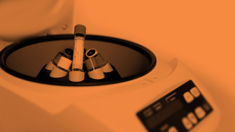
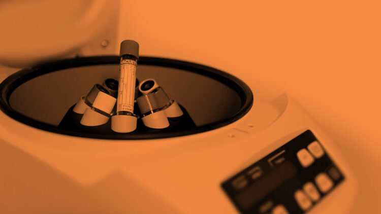
Platelet concentrates (PCs), meanwhile, are blood extracts, obtained after processing a whole blood sample through centrifugation (Schliephake et al 2018). Perhaps the best known of these in dentistry are Platelet-Rich Plasma (PRP) and Plasma Rich in Growth Factors (PRGF). Autologous PRP was first developed in the early 1970s, which means PCs have been used in modern medicine for more than four decades.
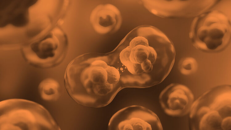
They are used due to their impact on tissue regeneration by facilitating angiogenesis and various additional phases during wound healing, including:
- Cell recruitment
- Proliferation
- Remodelling
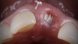
When no anticoagulant or thrombin/ calcium chloride is used and only one step of centrifugation is employed during which coagulation occurs, Platelet‐Rich Fibrin (PRF) can be prepared (Schliephake et al 2018). The coating of implants with PRF either before or during implant placement has demonstrated a meaningful increase in Implant Stability Quotient (ISQ) values. Additionally, the use of PRF matrices in conjunction with implant insertion to cover the bone before tissue closure has shown less bone loss during the first three months (0.35 mm). However, it is imperative to note that use of PRF during implant placement cannot yet be unreservedly recommended due to limited and premature clinical evidence (Schliephake et al 2018). Nevertheless, Leukocyte- and Platelet-Rich Fibrin (L-PRF®) offers a possible alternative.
When leukocytes and PRF are combined
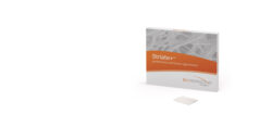
L-PRF is a 3D autogenous combination of PRF derived from the patient’s blood (Schliephake et al 2018). A simplified chairside procedure results in the production of a thin, compressed layer of platelet rich fibrin that is strong, pliable and suitable for suturing. This natural fibrin network is rich in platelets, growth factors and cytokines that are derived from the blood platelets and leukocytes (Schliephake et al 2018). The presence of these proteins has been reported to produce rapid healing, especially during the critical first seven days after placement (Dohan et al 2009). This network promotes more efficient cell migration and proliferation without chemical or bovine thrombin additives (Dohan et al 2006).
In a nutshell, L-PRF possesses the following properties:
- Simple and economic (Toffler et al 2009)
- Natural – 100% autologous (Toffler et al 2009)
- Thin fibrin matrix and plugs (Toffler et al 2009)
- Rich in leukocytes, platelets and fibrin (Dohan et al 2010)
- Slow release at ≥ _7 days (Dohan et al 2010)
- Matrix for bone graft material (Simonpieri et al 2012).
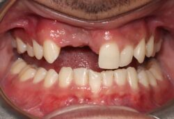
Clinically, L-PRF displays excellent working properties. This biomaterial is resilient, strong and pliable, making it easy to manipulate. It can be cut to size and is sufficiently supple to adapt to many anatomical areas, including but not limited to:
- Bone defects (Simonpieri et al 2012)
- Extraction sockets (Toffler et al 2009; Dohan et al 2010; Simonpieri et al 2012; Del Corso et al 2012)
- Sinus and dental ridge augmentation (Toffler et al 2009; Simonpieri et al 2012)
- Palatal defects (Jain et al 2012)
- Maxillary bone atrophy (Schliephake et al 2018; Simonpieri et al 2012).
L-PRF is adhesive in nature and very receptive to suturing. In addition, there is ample working time since L-PRF is stable at room temperature for several hours (Toffler et al 2009).
L-PRF in practice
Offering a practical application of L-PRF, the IntraSpin system establishes a three-step protocol for drawing and spinning the patient’s blood, removing the fibrin clot and processing it in the Xpression® fabrication kit. A thin, compressed layer of L-PRF or plugs for extraction sites can then be formed, using either the internal plate or the piston assembly.
L-PRF advantages include:
- No anticoagulant
- No bovine thrombin
- No heating
- No pipetting
- No second spin
- No chemical additives
- No expensive consumables
In addition, IntraSpin has been reported to be the most stable centrifuge and offers up to an 80% reduction in undesirable vibrations. Since centrifuge characteristics may affect the cellular quality, growth factors and fibrin architecture of an L-PRF clot and matrices, this is a key issue. Indeed, some other centrifuges tested did not produce PRF but PRF-like materials with a damaged and almost destroyed cell population when using the standard protocol developed initially for L-PRF. This meant it is not possible to classify these products in the L-PRF family (Dohan et al 2017).
With data suggesting a promising role for platelet concentrates in the clinical setting of wound healing and tissue regeneration and evidence supporting a strong pro-angiogenic effect of L-PRF in vitro and in vivo (Ratajczak et al 2018), the time has come to consider using L-PRF in your implant practice.
The L-PRF difference
- Cost-effective – Simple procedure with a low per patient cost
- Safe – Derived from the patient’s own blood, reducing the risk of disease transmission and allergic reaction or rejection
- Natural – 100% natural and concentrates the growth factors that are present in the patient’s own blood, allowing the patient’s body to heal itself naturally
- Healing properties – Promotes enhanced wound healing
- Simple procedure – FDA cleared
Readers can answer the questions and claim their free hour of enhanced CPD by completing the form at the end of this article or by emailing their answers to cpd@biohorizons.com.